
Endo-Diagnosis of Early NPL and
Indications for Resection Technique (RT)
Atlas of Endoscopy with Narrow Band Imaging
M Muto, K Yao, Y Sano (eds.), 1st JP ed., Nankodo Co Ltd, Tokyo, 2011
- CHAP 1: K Gono. Principles & History of NBI, pp 3-10.
- CHAP 2: M Muto, K Yao, Y Sano. Tips for obtaing optimum viewing with NBI, pp 11-30.
- NBI Atlas: Structure: Overview/ Normal/ Nonneoplastic/ Neoplasias for Pharynx & Esophagus, Stomach, Duodenum, Colorectum & Anal Canal, pp 33-340.
Atlas of Early Neoplasias of the Gastrointestinal Tract
Endoscopic Diagnosis and Therapeutic Decisions
F Berr, T Oyama, T Ponchon, N Yahagi (eds.), 2nd ed., SPRINGER, New York
- CHAP 1 F Berr, T Ponchon, T Uraoka. Endoscopic detection and analysis of mucosal neoplastic lesions, pp 3-24.
- CHAP 6: T Ponchon, F Berr, T Oyama. Endoscopic Screening & Surveillance, pp 101-118.
Endoscopic detection
with standard white light imaging (WLI on scope insertion in esophagus to colorectum), and NBI/BLI/i-scan TE in squamous cell mucosa (pharynx on scope insertion; esophagus on scope withdrawal).
Endoscopic analysis
- macroscopic type & signs on standard WLI with insufflation / desufflation
- microvascular pattern (V) on magnifying NBI / BLI / i-scan TE.
- microsurface pattern (S) on magnifying (≥60-80-fold) chromoendoscopy (algorithm) or magnifying NBI / BLI (m-NBI).
Endoscopic diagnosis based on these 3 criteria → predicted pT category & grading → indication for RT
Special diagnostic criteria: CHAP 8 (Barrett) & CHAP 12 (IBD) In: Atlas of Early Neoplasias of the GIT, Springer 2019
Lectures for self instructionsResection Techniques (RT) for EC and precursor NPL
Endoscopic resection techniques (ER)
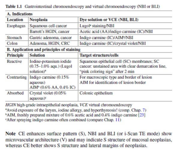
Resection Techniques Indications for en bloc ER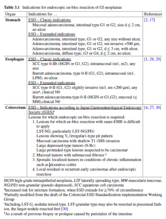
CHAP 3: T Oyama, N Yahagi. Principles of endoscopic resection (ER): Diagnostic and curative ER. In: Atlas of Early Neoplasias of the GIT, 2019, pp 47-53.
Surgical resection with lymphadenectomy (SR+LNE)
Syllabus ESD Hands-on Workshop - Berlin 2024, p26-30 (in downloads)
Histopathology of Early Mucosal Neoplasias - histologic carcinogenesis and curative resection (CR)
CR criteriaCHAP 2: D Neureiter, T Kiesslich. Histopathology of early mucosal neoplasias in the GIT.
In: Atlas of Early Neoplasias of the GIT, 2019, pp 25-45.
Lectures for Diagnostic Self-Instruction
- (2016) Ponchon T: How to take HQ-images for endoscopic staging
- (2018) Mitrakov A & Yao K: E-learning for endo-detection of early gastric cancer (Quiz)
- (2016) Maehata T: Endoscopic analysis of early colorectal neoplasias
- (2017) Uraoka T: Colorectum
- (2017) Takahashi A: Squamous epithelial Esophagus
- (2018) Takahashi A: Magnifying endoscopic analysis of early gastric cancer
Quizzes for Self-Instruction
- (2016) Oyama T: Detection and analysis of early neoplasia in columnar cell-lined upper GI tract
- (2017) Oyama T: Esophago-gastric lesions
- (2018) Maehata T: How to assess early colorectal neoplasias for resection (Quiz)
- (2017) Yahagi N: Colorectal lesions
References
- CHAP 7: Oyama T. Squamous cell-lined esophagus and hypopharynx: mucosal neoplasias. In: Atlas of Early Neoplasias of the GIT, 2019 pp 121-147.
- CHAP 8: Deprez PH, Toyonaga T. Columnar epithelium-lined (Barrett´s) esophagus: mucosal neoplasias. In: Atlas of Early Neoplasias of the GIT, 2019 pp 149-173.
- Oyama T, et al., Prediction of the invasion depth of superficial squamous cell carcinoma based on microvessel morphology: magnifying endoscopic classification of the Japan Esophageal Society. Esophagus 2017;14: 105-112. (open access paper)
- Goda K et al, Diagnostic utility of a novel magnifying endoscopic classification for superficial Barrett´s esophagus-related neoplasms: a nation-wide multicenter study. Esophagus 2021;18:713-723. (open access paper)
- Yao K, et al., Detection and characterization of early gastric cancer for curative endoscopic submucosal dissection. Dig Endosc 2013;25 Suppl 1: 44-54./li>
- Muto M, et al., Magnifying endoscopy simple diagnostic algorithm for early gastric cancer (MESDA-G). Dig Endosc 2016;28: 379-393
- CHAP 9: Oyama T. Stomach: Mucosal neoplasias. In: Atlas of Early Neoplasias of the GIT, 2019 pp 175-222.
- Sano Y et al, JNET classification. Dig Endosc 2016;28:526-33.
- CHAP 11: Wagner A, Maehata T, Berr F, Yahagi N. Colorectum: mucosal neoplasias. In: Atlas of Early Neoplasias of the GIT, 2019 pp 241-289.
- CHAP 12: Yahagi N, Maehata T, Nakayama A. Chronic inflammatory bowel disease in remission: mucosal neo-plasias. In: Atlas of Early Neoplasias of the GIT, 2019 pp 291-305.