Microsurface patterns (S) of NPL in the GIT
Analysis of microsurface pattern best is performed with magnifying CE (mCE, >60x). Chromoendoscopy uses different dye techniques, depending on epithelium and localization in the GIT, crystal violet only for targeted CE (<2 mL) (see Table 1.1 & chap 1, pp 5-6.):
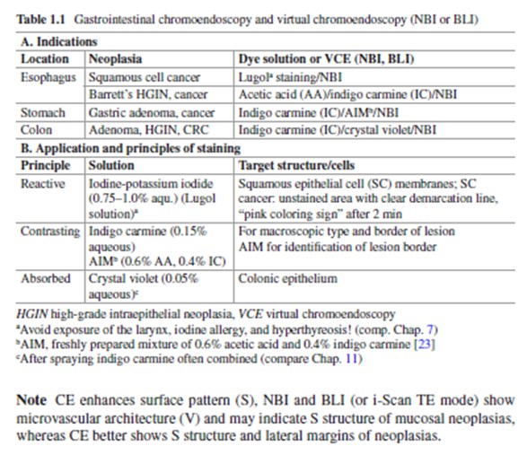
S/V analysis of NPL in SC-esophagus, Barret´s esophagus, Stomach and Duodenum, and IBD-Colorectum see chap.s 7 – 10, 11; Atlas of Early Neoplasias of the GIT,2nd ed., Springer New York, 2019
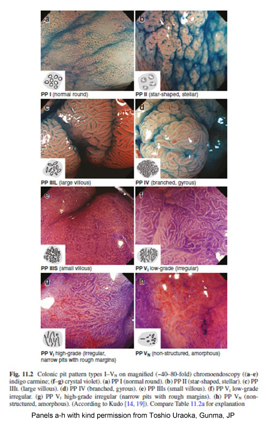
Colorectum - Kudo´s Pit Pattern
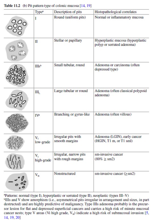
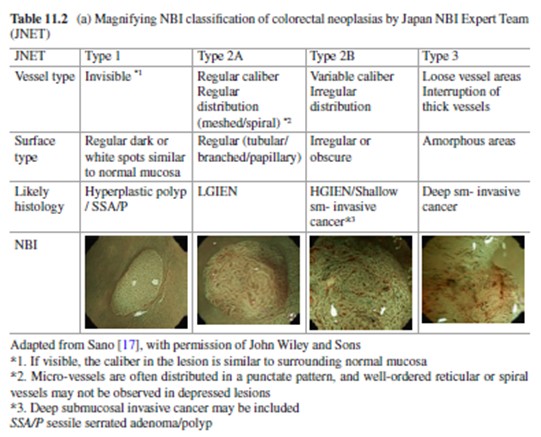
Colorectum – Kudo´s Pit Pattern (for explanation compare Tab. 11.2)