Microvascular patterns (V) of NPL in the GIT
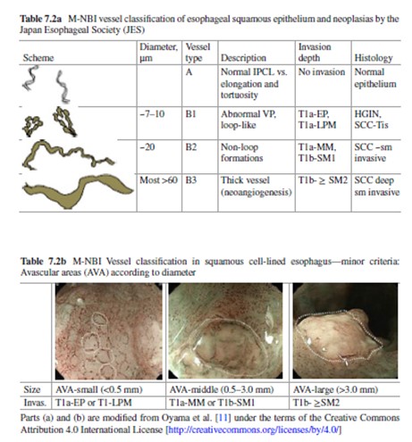
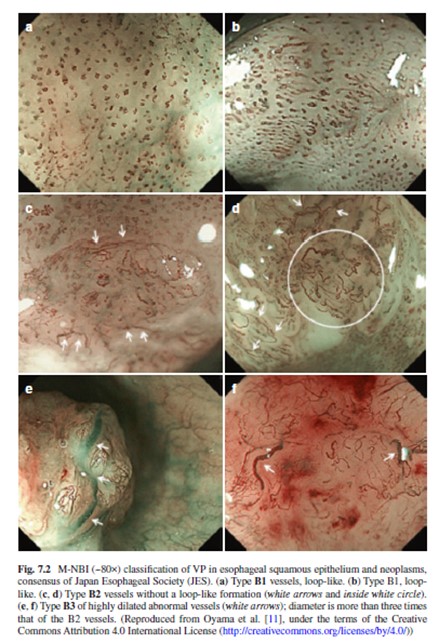
Squamous cell-lined esophagus
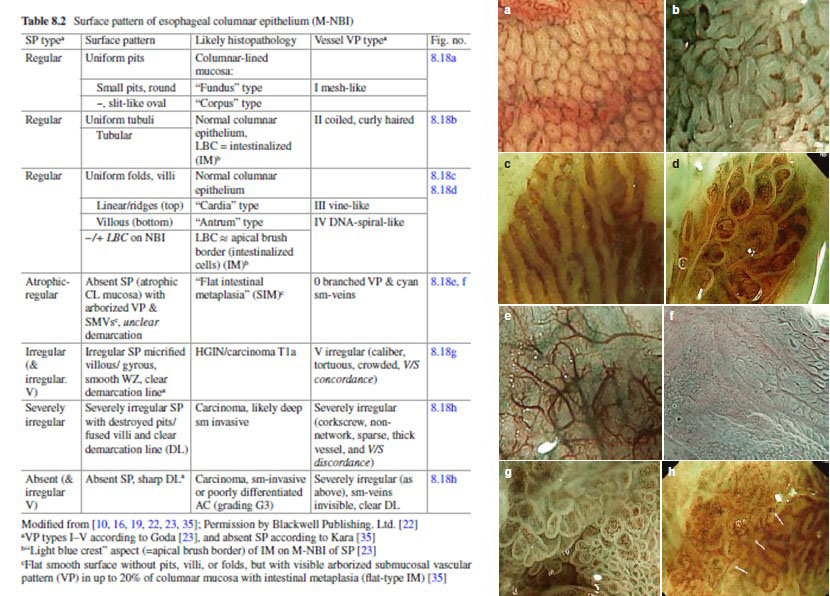
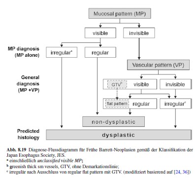
Barrett´s esophagus (cylinderepithelial metaplasia)
From: chap 8, Deprez PH, Toyonaga T. Barrett´s esophagus: Mucosal Neoplasias. In: Atlas of Early NPL of the GIT. 2nd ed., Springer New York, 2019.
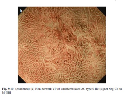
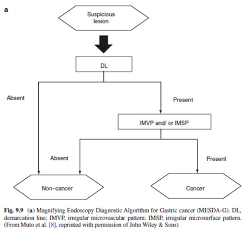
Stomach (Fundus ~ pit pattern [ V = network], Antropylorus ~ villous pattern [ V = spiral])
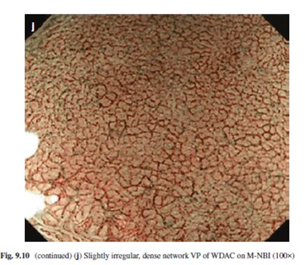
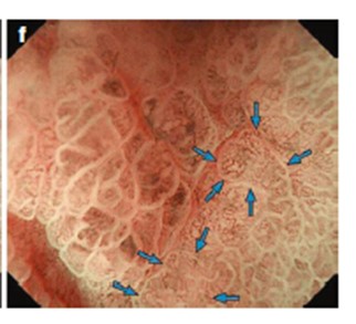
From Chap. 9 Oyama T. Stomach: Mucosal NPL, Atlas of Early Neoplasias of the GIT, Springer New York 2019
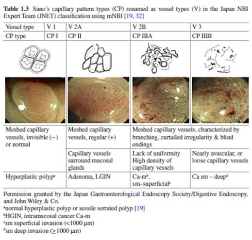
Colorectum ( V types 1-3 of JNET classification)